Haemodynamic response, neurovascular coupling, and measuring brain activity indirectly
When you work out, your body supplies nutrients (such as oxygen) to your muscles by adjusting the blood flow. The haemodynamic response is the name of this biological process that quickly delivers blood to active brain tissue. Basically, this biological fact states also that a pumped-up bicep is going to need a release of oxygen to it through the blood flow.
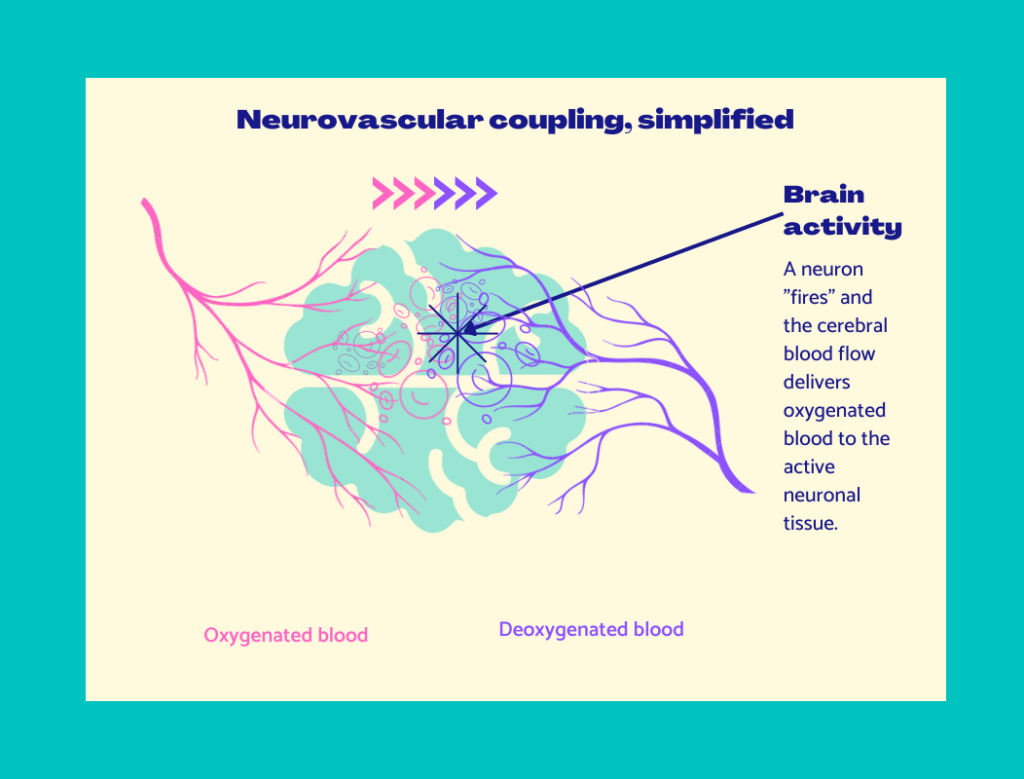
Similarly, when your brain is performing a specific task, the area that processes the task will first receive oxygenated blood. The active neuronal tissue “consumes” the oxygen from the blood flow, after which the brain area’s blood circulation will have more deoxygenated blood. This connection between neuronal activity and blood flow is also referred to as neurovascular coupling.
While some imaging tools measure brain activity directly, hemodynamic brain imaging tools measure brain activation indirectly by measuring the blood flow connected to the neurons. For instance, functional magnetic resonance imaging (fMRI) is a big scanner that measures brain activity extremely accurately. Compared to an expensive fMRI scanner that can hardly be used in home settings, fNIRS is cheap and portable. Both are functional imaging methods, meaning they measure brain activity relating to a certain brain process safely without stimulating neural activity. They measure brain activity indirectly by measuring the hemodynamic response related to brain activity through the mechanism of neurovascular coupling.
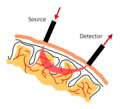
Moreover, fNIRS measures the blood flow in the brain using so-called optodes emitting and detecting near-infrared (NIR) lights. NIR light passes safely through biological tissue and illuminates it a few centimetres deep. There it detects oxygenated and deoxygenated blood. Hence, fNIRS can measure changes in oxygenated and deoxygenated blood. The darker the tissue, the harder it is to measure brain activity due to the absorption of the NIR light.